Headline News
HYPOMYELINATING LEUKODYSTROPHIES — UNRAVELLING MYELIN BIOLOGY
HYPOMYELINATING LEUKODYSTROPHIES — UNRAVELLING MYELIN BIOLOGY
Wolf NI, Ffrench-Constant C, van der Knaap MS
https://www.nature.com/articles/s41582-020-00432-1
 Dr. van der Knaap and colleagues recently published an update on hypomyelinating leukodystrophies! This comprehensive review emphasizes the latest advances in myelin biology and genetic underpinnings of these disorders.
Highlights include:
- Myelination is a complex process that involves interactions between oligodendrocytes, axons, astrocytes, and microglia.
- Myelin has an essential role in nurturing axons. Absence of this axonal support accounts for the gradual clinical decline in patients with hypomyelination.
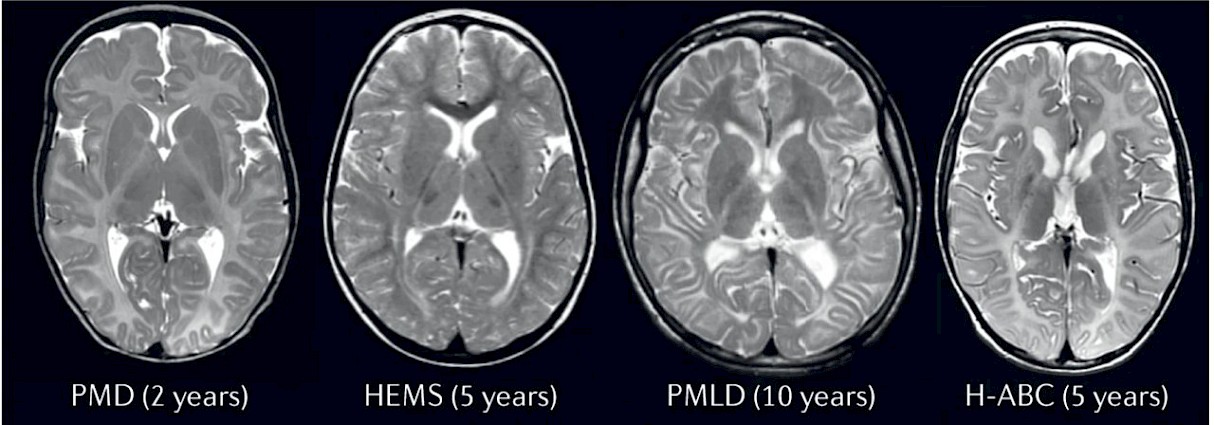
- Myelination occurs in an expected temporal and spatial pattern, which can be assessed on MRI. When disordered, pattern recognition by MRI can provide a clinical diagnosis in many patients.
- Radiological techniques can quantify myelin e.g. T1 mapping, multicomponent T2 relaxation imaging, mcDESPOT, g ratio, quantitative diffusion parameters, and NODDI. The best estimates of myelin quantity and quality are likely to come from combining multiple techniques. These quantitative MRI techniques can provide measures of white matter myelin content, with potential to act as biomarkers.
- Genes associated with hypomyelination include those that encode structural myelin, those that encode the proteins involved in RNA translation, as well as some lysosomal proteins.
Development of adequate treatment strategies for hypomyelinating leukodystrophies relies upon knowledge of the natural disease evolution on conventional brain MRI, an understanding of novel MRI techniques and their ability to quantify and characterize myelin, as well as close collaboration with clinicians and myelin biologists. Global collaboration is essential given the rarity of the disorders!
Arzu Ozturk, MD
UC Davis Health